

Could a cam deformity on your scan be unrelated to your hip pain? It can be unsettling to find an unfamiliar term in an imaging report, especially if you’re worried that your joint is being damaged. But a cam-shaped area of bone doesn’t automatically mean you have painful hip impingement or that the problem will inevitably get worse.
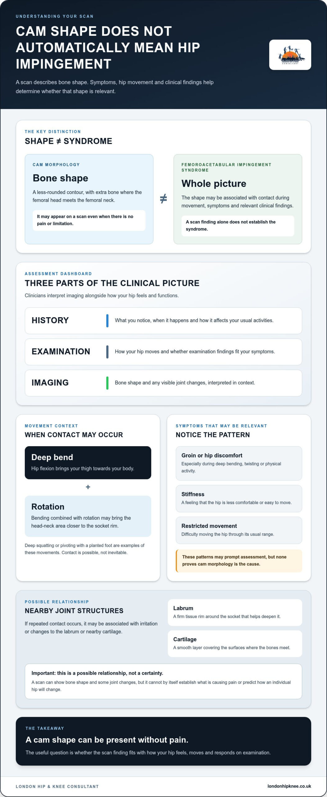
The important distinction is between cam morphology, the shape seen on a scan, and femoroacetabular impingement, which may occur when that shape causes contact in the hip and fits with your symptoms. Many people have cam morphology without pain. If you’re experiencing groin discomfort, stiffness or restricted movement, clinicians consider your history and examination alongside imaging rather than relying on the scan alone.
This guide explains what a cam deformity means, which symptoms may be relevant and how an assessment is made. You’ll also learn about possible next steps, from adjusting activities and physiotherapy to when hip arthroscopy might be considered. Treatment depends on how the whole picture fits together, not on one scan finding alone.
Key Takeaways
- A cam deformity describes a change in the shape of the femoral head and neck. Its presence on a scan doesn’t, by itself, explain pain.
- Notice when symptoms such as groin discomfort or restricted movement occur, particularly during hip flexion or rotation, and share these details during assessment.
- Scan findings need to be considered alongside your symptoms, hip examination and any changes to nearby joint structures.
- Clinicians review your history, examine your hip and interpret relevant imaging before discussing next steps.
- If ongoing hip symptoms affect daily activities, prepare for an appointment by bringing scan reports and noting how your symptoms affect you.
What is cam deformity, and does it always cause hip pain?
Seeing “cam deformity” on an X-ray or scan report can raise an immediate worry: does this explain your hip pain, or mean the joint is being damaged? Not necessarily. The term describes the shape of part of the hip bone. On its own, it doesn’t confirm that you have a painful hip condition or that treatment is needed.
Where is the cam shape, and what does it describe?
Your hip is a ball-and-socket joint. The ball, called the femoral head, sits in the socket of the pelvis and joins the thigh bone through a narrower section called the femoral neck. In a typical hip, the transition between the head and neck is relatively smooth. With cam morphology, the contour is less rounded, with extra bone at this junction.
In short, a cam deformity describes an altered bone contour where the femoral head meets the neck. It describes shape, rather than a separate growth or tumour. Imaging may show this shape even if you have no pain or limitations.
Cam morphology versus femoroacetabular impingement
The shape alone is different from femoroacetabular impingement syndrome. During certain movements, particularly bending the hip deeply or rotating it, the femoral head-neck area may come into contact with the edge of the socket. In some people, this contact is associated with symptoms and clinical findings. In others, the same shape causes no noticeable problem.
Clinicians use the term femoroacetabular impingement syndrome when the overall picture supports it, rather than simply because a scan shows cam morphology. They consider symptoms and examination findings alongside imaging. Groin pain, for example, can have different causes, so one symptom or scan description cannot establish the diagnosis by itself. A Femoroacetabular impingement overview explains the broader condition, but your own scan needs to be interpreted in the context of your hip and symptoms.
The key point: a cam shape can be present without pain. If your report mentions it, the next question is not just what the bone looks like, but whether the finding fits with how your hip feels and moves. That distinction helps avoid treating an imaging finding as a diagnosis on its own.
How can cam morphology affect hip movement and joint structures?
A cam-shaped area matters because of where it sits: at the junction between the ball of the hip and the thigh bone. Its effect, if any, depends on how the bones meet during movement and whether this corresponds with symptoms or findings on examination. The shape alone doesn’t show that damage has occurred.
What happens during hip flexion and rotation?
Flexion means bending your hip, bringing your thigh towards your body. Add rotation, and the rounded head-neck junction may move closer to the rim of the socket. This combination can happen in a deep squat or when you pivot with your foot planted. If the contours meet, contact may occur sooner or differently than in a hip with another shape.
That doesn’t mean squatting, turning or other everyday movements inevitably harm your hip. Range of motion, bone shape, activity and sensitivity to movement vary between people. A position that feels uncomfortable for one person may be entirely comfortable for another. Symptoms can also have causes unrelated to cam morphology, so movement-related pain needs to be understood in context.
How may the labrum and cartilage be involved?
The acetabulum is the socket in the pelvis. Around its edge sits the labrum, a rim of firm tissue that helps deepen the socket. A smooth layer of cartilage covers the surfaces where the bones meet. If repeated contact occurs, it may be associated with irritation or changes to the labrum or nearby cartilage. This is a possible relationship, not a certainty for every hip.
A scan may help show bone shape and some changes in the joint, but it cannot by itself establish what is causing pain or predict how an individual hip will change. The Femoroacetabular Impingement Syndrome resource outlines imaging features. These still need to be interpreted alongside symptoms and clinical examination.
The reasons cam morphology develops aren’t fully explained, and a scan finding alone can’t tell you why it formed. If hip discomfort limits movement or affects your usual activities, a specialist assessment can help relate your symptoms to the examination and imaging. You can read more about specialist hip care in London.
Cam deformity myths: what symptoms and scans can, and cannot, tell you
A scan report can make a finding sound like a diagnosis, but it isn’t the whole story. A cam deformity does not automatically cause pain, mean the hip is being damaged or indicate that an operation is needed. Some people have this bone shape without symptoms. Treatment decisions depend on how your hip feels and functions, what an examination shows and whether the imaging findings fit that picture.
Which symptoms may prompt an assessment?
Hip or groin discomfort during deep bending, twisting or physical activity can be a reason to seek assessment. Some people also notice stiffness or difficulty moving the hip through its usual range. These patterns may be relevant, but none proves that cam morphology is the cause.
Similar symptoms can arise from other hip conditions, and pain location alone can’t identify the source. Rather than diagnosing yourself from an online symptom list, note when discomfort occurs, what movements bring it on and how it affects daily activities. This history helps a clinician consider possible explanations and decide whether further assessment is appropriate.
What can an X-ray or MRI show?
An X-ray can help show the shape of the bones around the hip. MRI can provide information about soft tissues, including the labrum and cartilage, as well as other structures in the area. Each scan offers different information, and which imaging is useful depends on the clinical question.
A scan finding needs to be read alongside your symptoms and the results of a physical examination. For instance, a bone shape seen on an X-ray may not explain discomfort if your symptoms and examination point elsewhere. Equally, the absence of a particular finding on one image doesn’t replace a full clinical assessment. No single scan determines that surgery is necessary.
Seek medical advice if hip symptoms persist, worsen or interfere with walking and everyday activities. Severe, sudden pain, an inability to bear weight, or pain accompanied by fever or feeling unwell warrants prompt medical assessment through appropriate NHS services. These symptoms shouldn’t be assumed to be caused by a cam shape. Timely assessment can help establish what may be happening and what to do next.
What happens after a cam deformity diagnosis?
A scan finding is a starting point for discussion, not a treatment plan. Your clinician will build a clearer picture by asking about your symptoms and the activities that affect them, examining hip movement and function, and reviewing relevant imaging alongside those findings. Together, you can discuss what the evidence suggests and agree on a suitable next step.
The decision depends on more than bone shape. Your level of discomfort, how it affects walking or other activities, examination findings and any associated changes in the joint all matter. If symptoms are mild or absent, monitoring or no specific treatment may be appropriate. If symptoms affect your daily life, options can be considered in light of your individual circumstances.
What may non-surgical management involve?
For some people, an initial approach may include adjusting activities that repeatedly bring on discomfort and working with a physiotherapist on movement, control and strength. The aim is to find a manageable way to stay active whilst addressing difficulties with hip function. Advice should be tailored to you. There isn’t one set of exercises or activity changes that suits everyone.
Reviewing symptoms over time can help assess whether an approach is useful and whether the plan needs to change. If you’re considering pain relief or other treatment, discuss what is appropriate with a clinician who understands your medical circumstances.
When might hip arthroscopy be considered?
Hip arthroscopy is keyhole surgery that allows a surgeon to assess and treat selected problems inside the joint. It may be discussed for some people whose symptoms and clinical findings fit with hip impingement, particularly when non-surgical measures haven’t adequately addressed their difficulties. A cam shape on an X-ray alone is not a reason to proceed.
A specialist will consider whether the likely benefits justify the limitations and risks of surgery in your particular case, including any other joint findings. The discussion should help you understand the alternatives and what an operation may involve, so that any decision is informed and individual.
London Hip & Knee Consultant provides hip arthroscopy for selected cases. If ongoing hip symptoms affect your movement, you can arrange a specialist hip assessment to discuss your symptoms and imaging with a clinician.
When should you seek specialist advice about cam deformity in London?
Consider an assessment if hip symptoms persist or begin to affect walking, sleep, exercise or everyday tasks. A scan finding may be worrying, but an appointment can help put it into context. A clinician can discuss what you’re experiencing, examine how your hip moves and review relevant imaging before talking through possible next steps. This doesn’t assume that the scan finding is the cause or that you’ll need treatment.
How to prepare for a hip assessment
A few notes can help you explain what has changed and make the conversation more useful. Before your appointment, consider recording:
- When the discomfort began and whether it has changed over time.
- Which movements or activities aggravate it, and what makes it easier.
- How it affects walking, sleep, work, exercise or other usual activities.
- Any previous treatment, physiotherapy or rehabilitation, and how you responded.
Bring relevant imaging reports and details of previous scans. You can also note questions you’d like answered, such as what the findings may mean, which options could be appropriate, and what recovery or potential risks might involve if treatment is considered. You don’t need to interpret the images yourself.
How a specialist assessment can clarify your options
Mr Parag Jaiswal is a Consultant Orthopaedic Surgeon with a specialist focus on adult hip and knee conditions. An assessment can consider your symptom history, examination and imaging together, helping distinguish an incidental bone-shape finding from a problem that may relate to your symptoms. The discussion may include non-surgical approaches. Hip arthroscopy is considered for selected cases after individual assessment, not simply because a scan shows a cam deformity.
If persistent symptoms are limiting your usual activities, an assessment is a practical way to discuss your concerns and possible next steps. Arrange a specialist hip assessment in London.
Take the next step towards understanding your hip
A cam deformity describes bone shape, but it doesn’t automatically explain hip pain or mean that treatment is needed. The key is whether your symptoms, examination and imaging findings fit together. Persistent discomfort or restricted movement deserves attention. Understanding what affects your hip can help you make informed choices about what to do next.
At a specialist assessment, Mr Parag Jaiswal, Consultant Orthopaedic Surgeon, can review your symptoms and relevant imaging before discussing options. These may include non-surgical care. Hip arthroscopy may be considered for selected cases, but it isn’t an automatic response to a scan finding.
If hip symptoms are affecting your daily life, take a considered next step and arrange a specialist hip assessment in London.
Frequently Asked Questions
Is a cam deformity always painful?
No, a cam shape can be present without causing symptoms. Whether it is relevant to your hip depends on how your symptoms, physical examination and imaging findings fit together. Hip pain can have other causes, so a scan description alone can’t confirm femoroacetabular impingement syndrome. If discomfort continues or starts to affect your everyday activities, discuss it with a qualified clinician who can assess the whole picture.
Can a cam deformity get worse over time?
A cam deformity describes bone shape, but it doesn’t mean that symptoms or joint changes will inevitably progress. What happens can differ between individuals, and a scan cannot predict your personal course on its own. If pain, stiffness or difficulty with activities changes, a clinician can review your symptoms and relevant imaging. This is more useful than assuming that the finding means your hip is getting worse.
What does cam deformity feel like?
Some people feel nothing at all. If symptoms occur, they may include discomfort in the groin, reduced hip movement or pain during activities involving deep bending or rotation. These symptoms aren’t specific to cam morphology and can have other causes. Rather than relying on pain location or an online symptom list to self-diagnose, note when discomfort occurs and how it affects your function, then discuss this during an assessment.
How is cam deformity diagnosed?
Assessment usually brings together your symptom history, a physical examination and imaging when appropriate. An X-ray may show the shape of the bones, while an MRI can help assess soft tissues such as the labrum. Imaging needs to be interpreted alongside your symptoms and examination findings. A scan alone can’t establish that the bone shape is causing pain or determine that surgery is needed.
Does cam deformity always need surgery?
No. A cam shape on imaging doesn’t automatically need treatment, and surgery isn’t decided by the scan alone. Depending on symptoms and clinical findings, options may include adjusting activities or physiotherapy. Hip arthroscopy may be discussed for selected people after specialist assessment. A clinician should explain the potential benefits, limitations and risks in relation to your individual hip and circumstances before you consider an operation.
Can physiotherapy help with cam deformity?
Physiotherapy may help some people manage symptoms, build strength or adapt how they move, but it doesn’t alter the shape of the bone. Whether it’s suitable depends on your symptoms, examination and goals. A clinician or physiotherapist can help shape an appropriate rehabilitation plan and review how you respond over time. Avoid assuming that one set of exercises is right for every hip or symptom pattern.
When should I see a hip specialist about cam deformity?
Consider an assessment if hip symptoms persist, restrict everyday activities or fail to improve with initial self-management. Seek prompt medical advice for severe or sudden symptoms. Before an appointment, note which movements trigger discomfort and bring relevant scan reports or previous imaging. A specialist can review your symptoms, examination and imaging together, then discuss whether the cam finding may be relevant and what options could suit your situation.